Misr Lab for Medical Analysis in Giza – Your Health Services Since 2005
Misr Lab for Medical Analysis is your trusted destination for medical tests in Giza. Moreover, we offer comprehensive analyses with accurate and fast results. In addition, we provide a home sample collection service for your utmost convenience. Therefore, do not hesitate to contact us via WhatsApp and book your appointment now.
Thursday – Saturday
10:00 – 22:00
01013330947
Misr Lab
Comprehensive Medical Analysis
Discover Our Clinical Health Experiences
Knowing more about yourself means finding the answers you need in health.


Diagnostic Excellence
Science and Technology at Your Service
Speed and Accuracy
Clear and Reliable Diagnosis
Top Professionals
Experience That Makes a Difference
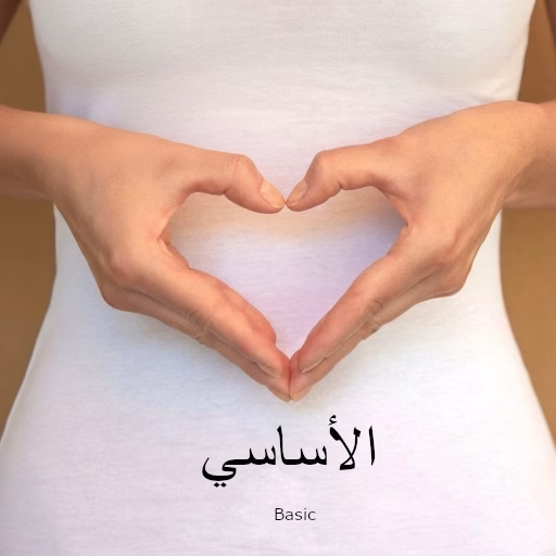
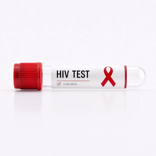
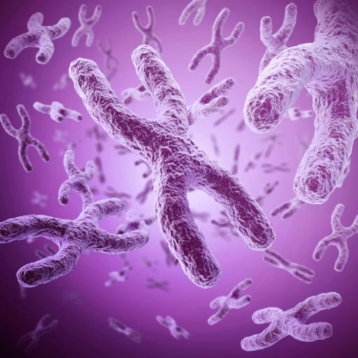
Nutrition · Genetics · Early Detection · Sexual Health · Fertility & Pregnancy · SportsNutrition · Genetics · Early Detection · Sexual Health · Fertility & Pregnancy · Sports
Discover Our Clinical Health Experiences


Catalog featuring over 4,000 specialized reference tests • Advanced technology • Over 20 years of experience • Innovation at your service • Certified qualityCatalog featuring over 4,000 specialized reference tests • Advanced technology • Over 20 years of experience • Innovation at your service • Certified quality
[fusion_widget type=”Fusion_Widget_Facebook_Page” margin_top=”” margin_right=”” margin_bottom=”” margin_left=”” fusion_widget_facebook_page__title=”Find us on Facebook” fusion_widget_facebook_page__page_url=”https://www.facebook.com/1031429916952409″ fusion_widget_facebook_page__width=”500″ fusion_widget_facebook_page__show_faces=”on” fusion_widget_facebook_page__show_stream=”on” fusion_widget_facebook_page__show_events=”off” fusion_widget_facebook_page__show_messages=”off” fusion_widget_facebook_page__small_header=”on” hide_on_mobile=”small-visibility,medium-visibility,large-visibility” fusion_display_title=”no” fusion_border_size=”0″ fusion_border_style=”solid” fusion_widget_facebook_page__show_header=”off” fusion_bg_color=”rgba(0,0,0,0.01)” /]
Book Your Analysis Now in Giza
Choose the right package for you and we’ll do the rest. Home collection service available. Therefore, don’t hesitate to contact us now.
Misr Lab
???? 26 King Faisal Street – 4th Floor – Giza 128
???? 39 Al-Ahram Street – 3rd Floor – Giza
????⚕️ Dr. Mohamed Saleh
???? +20 233903528 | +20 01013330947
✉️ info@misrlab.com | ???? www.misrlab.com
“`